
Renal Resistive Index Calculator
This calculator is used to compute the renal resistive index, a measure of renal artery resistance that may help in the diagnosis of renal vascular hypertension.
Inputs
Result
Enter values to calculate
Formula
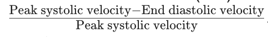
RRI = (Peak Systolic Velocity - Peak Diastolic Velocity) / Peak Systolic Velocity Calculator Diagram
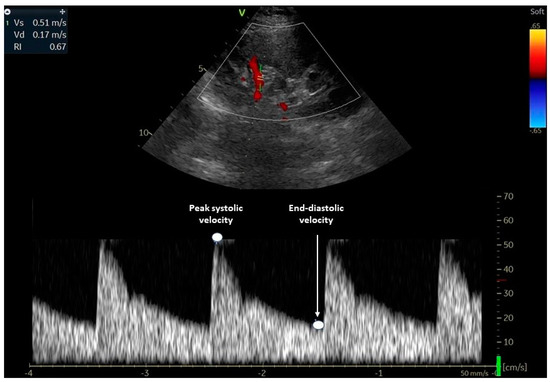
Renal resistive index (RRI) is calculated using pulse wave doppler over the renal artery within the renal cortex.
Theory and Practice
Renal resistive index is an assessment of the peak systolic and end diastolic flow through the renal artery at the level of the renal cortex. In critically ill and trauma patients, it is a reliable predictor of acute kidney injury. In shock, the kidney is often the first organ that we see signs of malperfusion in, often manifesting as low urine output.
Renal arteries are typically low resistance, with continuous forward flow throughout systole and diastole. An elevated renal resistive index indicates a state of increased resistance within the renal vasculature, which may be due to systemic and intra-renal factors. Systemic factors include changes in MAP, venous congestion, and central venous pressure, while renal factors include tubular injury (ATN, glomerulonephritis) and microvascular dysfunction.
A normal value for renal resistive index is <0.7, with values >0.7 predictive of acute kidney injury.
Consider both systemic and renal factors when evaluating patients with shock and elevated resistive indices. Are systemic contributors like venous congestion, bradycardia, or wide pulse pressure (Pulse Pressure Calculator) present? Is there other evidence of renal disease?
References
- 1. Lintner Rivera M, Prager R, Gushu MB, June S, Phiri T, Salameh JP, Johnson HC, Taylor T, O'Brien NF. Point-of-care Ultrasound to Assess Hemodynamic Contributors to Acute Kidney Injury in Pediatric Patients With Cerebral Malaria: A Pilot Study. Pediatr Infect Dis J. 2023 Oct 1;42(10):844-850. doi: 10.1097/INF.0000000000004021. Epub 2023 Jul 6. PMID: 37409812.
- 2. Turk, M., Koratala, A., Robertson, T., Kalagara, H. K. P., Bronshteyn, Y. S. Demystifying Venous Excess Ultrasound (VExUS): Image Acquisition and Interpretation. J. Vis. Exp. (219), e68107, doi:10.3791/68107 (2025).
- 3. Husain-Syed F, Birk HW, Ronco C, Schörmann T, Tello K, Richter MJ, Wilhelm J, Sommer N, Steyerberg E, Bauer P, Walmrath HD, Seeger W, McCullough PA, Gall H, Ghofrani HA. Doppler-Derived Renal Venous Stasis Index in the Prognosis of Right Heart Failure. J Am Heart Assoc. 2019 Nov 5;8(21):e013584. doi: 10.1161/JAHA.119.013584. Epub 2019 Oct 19. PMID: 31630601; PMCID: PMC6898799.
Contributors

Dr. Ross Prager

Isaac Bonisteel